Hand surgery

2main

This pathology of the hand is one of the most frequent and most unrecognized. It reflects a conflict between the deep flexor tendon and a digital pulley (pulley A1).

This conflict causes pain when fingers block in flexion. The blockage is due to the inflame at the level of the tendon causing a pull when passing through the pulley.

There are three stages of inctreasing severity :

  • Stage1 : Isolated pain at the base of the finger, no blockage is felt
  • Stage 2 : painful, sporadic blockage, mainly in the morning
  • Stage 3 : permanent blockage requiring the other hand to « have the finger back to its position ».
Manos

Generalities :

  • A distinction between Trapezo-metacarpal osteoarthritis (osteoarthritis) and scapho-trapezo-trapezoid arthrosis should be made
  • Osteoarthritis at the base of the thumb and the osteoarthritis scapho trapezo trapezoid should be classified on X-rays (Kapandi incidences)

The Dell’s radiological criteria for classification
Dell1 : Pinching or subchondral condensation with no subluxation, no osteophyte
Dell2 : Small osteophytosis and greater than one-third subluxation of the M1.
Dell3 : Osteophytosis with major pinching of the joint line and greater than one third subluxation of the metacarpal
Dell4 : Total destruction of the joint line with prominent osteophytes, geodes and trapezo-metacarpal ankylosis

Crosby’s radiological classification (STT)
Crosby0 : no apparent anomaly
Crosby1 : Decrease in half of normal joint space
Crosby2 : the joint line is barely visible
Crosby3 : Presence of erosions, sclerosis and irregularity of joint spacing

We have to determine the ground on which osteoarthritis of the base of the thumb occurs. This will determine the most appropriate treatment for the patient. The elements to be taken into account :

  • Age
  • Professional and sports activity
  • The dominant side
  • Theight of the trapeze
  • TM and STT
  • In all cases, it is imperative to try the medical treatmen tbefore the surgical treatment.
  • The medical treatment includes :
  • Short orthesis (TM + MP) diurnal
  • Long orthesis (wrist + STT + TM + MP) nocturnal
  • Infiltrations of Antalgic corticosteroids – delayed
  • Physiotherapy/ Kinesitherapy stabilizing TM without joint stress
  • For rhizarthrosis, it is necessary to explain the advantages and disadvantages of each technique

Trapeziectomy
Mobility ++ , Strength +++ , Postoperative Recovery Time 3 months, definitive TTT. At the same time, it cure the ST, any patient, dominant hand, Need for strength, associated STT, all sizes, Dell = all

mains3

What is Dupuytren’s contracture ?

Dupuytren’s disease or contracture is related to thickening of a fibrous structure under the skin of the palm of the hand and fingers

This thickening is accompanied by retraction, which limits the extension of the fingers..

Usually, the retraction of the fingers is not painful.

Very often another family member also has this disease (father, grandfather, uncle.

Alcohol and some drugs may promote development of Dupuytren’s disease.

When should you consider treatment?

Treatment should only be considered if the retraction prevents full extension of the fingers.

There is a very simple test: THE TABLE TEST

If one or more fingers cannot be extended enough to lay your hand flat on the table, the test is positive and surgical treatment is probably required.

If your test is negative, monitor the evolution.

The more the finger is retracted before the operation, the more difficult it is to restore full extension.

What are the surgical options?

Treatment involves removing the retracted tissue causing the disease. This is a delicate surgery because vessels and nerves are entangled with the Dupuytren’s bands.

In case of preoperative severe finger retraction, it is not always possible to restore full finger extension.

Sometimes because of skin defect, the palm of the hand is left open. Healing is then obtained with regular dressings for 2-3 weeks.

In almost all cases, the procedure is performed under local anaesthesia. You will leave the clinic the same day or the day after surgery. A skin graft may be used to cover skin defects or in case of recurrence.

This graft will most often be taken at the expense of the anesthetized limb (forearm or arm) leaving a filiform scar.

Nevertheless, a common behaviour is to be followed to ensure the best care from the various caregivers ( physician, nurse, physiotherapist, orthotist …).

Post-operative care : immobilization

An immobilization of the hand may be required depending on the degree of postoperative outcome and the ability to move the fingers freely to prevent recurrence of the flexion contracture.

Immobilization aims to improve healing in the most appropriate position and avoid scar bridles.

Physiotherapy :

Physiotherapy is often useful after an initial phase of immobilization to recover.

It helps for the drainage, perform an analgesic physiotherapy, provide a massage to the scars and soften them, recover passive and active mobility and if needed with the wearing of an orthesis.

Drugs :

Medical prescription will be made according to your age, your condition, your medical history, your possible allergic profile, and your ongoing treatment.

Analgesics are prescribed systematically. Postoperative antibiotics are generally not necessary.

douleur-au-coude-sd

What is compression of the ulnar nerve at the elbow ?

In the normal state, the ulnar nerve passes through a pseudo-canal at the elbow. Any limitation of the ulnar nerve motion will cause abnormal traction, which causes the nerve to suffer.

The dynamic anatomy of the parietal elbow during the motion from extension to flexion is partly responsible for the suffering of the ulnar nerve in the long course.

Finally, the chronic instability of the ulnar nerve may be responsible for nerve suffering, by a repetitive conflict with the medial epicondyle.

The usual compressions of the ulnar nerve occurs at the fibrous muscular arch, or Osborne fascia. This fascia is located at the end of the side which corresponds to a thickening of the envelopes of the anterior ulnar muscle.

What are the clinical signs ?

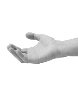
Paresthesias in or numbness on the palmar side , the 4th and 5th fingers are the most common motives behind consultation. They may be associated with a weakness of the hand.

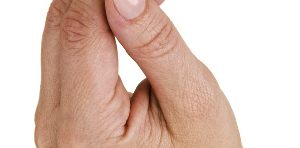
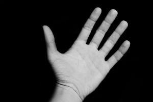
The clinician will then seek the set of sought clinical signs that reflect motor impairment: the Wartenberg sign which corresponds to the impossibility of bringing the little finger in contact with the other fingers (permanent abduction of the 5th finger), the Froment’s sign (weakness of the adductor and the deep bundle of the flexor), a muscular loss of the first interosseous space, a flattening of the hand with intermetacarpal atrophy of claws of the 4th and 5th fingers that can or can’t be reduced .

The clinical examination will end by seeking a chronic symptoms of ulnar nerve dislocation : from flexion to extension, the ulnar nerve is dislocated at the epitrochleo-olecranon gutter and passes forward of the medial epicondyle. This phenomenon is visible and palpable.

What other additional examination that should I have ?

An electromyogram that confirms the impaired sensory conduction and the slowing of the motor conduction at the elbow and an alteration of the recordings of the intrinsic muscles.

Simple x-rays of the elbow that can be used to diagnose bone damage.

What is the treatment ?

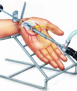
The treatment usually requires a surgy. It can be performed as an outpatient or during a short hospital stay of 1 to 2 nights, under loco-regional anesthesia.

The aim of the treatment is to perform a ulnar nerve decompression (neurolysis) by removing the causes of compression.

An anterior transposition of the nerve is also performed in the advanced motions. In case of transposition, the arm is immobilized in a splint for ten days.

There is a quick recovery of the sensory forms with almost immediate disappearance of paresthesias.

In acute forms of compression accompanied by hand amyotrophy, the disappearance of tingling is slower, and motion recovery takes several months.

Post-operative :

The neurolysis of the ulnar nerve is a surgey aiming to release the nerve from the arches compressing it at the level of the elbow.

The surgery allows at least to stop the evolution of paralysis and the loss of sensitivity, at best it allows the neurological recovery, and thus recovery of the contraction of the hand muscle and the sensitivity of the ring finger and the little finger.

The surgical treatment of the
ulnar nerve compression requires a fairly long recovery time (3 to 12 months).

The final result of decompression can not be assessed before one year after surgery, time required for the nerve to regenerate.

There two types of surgeries :

In Situ neurolysis :
Pure sensitive expression of the compression with stable nerve in its groove.

Often no immobilization is required.

Suitable analgesics are prescribed. A sick leave is often necessary for a few weeks.

A nurse should make your dressing every 4 days.

The first post-operative medical examination occurs at the end of the first week.

Neurolysis/ anterior subcutaneous transportation

Electromyographic motion expression or clinical expression or sensory expression on an unstable nerve or an impaired axis of the elbow.

An elbow splint is made in position of motion during the period of of healing (15 days).

Suitable analgesics are prescribed. A sick leave is often necessary for a few weeks.

Physiotherapy sessions may be required to overcome the motion deficit associated with the history of the nerve compression, the time for muscle recovery.

Pain in the joints of the hands on a gray background. Carpal tunnel syndrome. Care of male hands

What is Carpal tunnel Syndrome ?
1421454417_education

Carpal tunnel syndrome occurs when the median nerve of the hand is compressed within the transverse carpal ligament at the wrist.

The median nerve provides sensation to the thumb, the index finger, the middle finger and some parts of the ring finger .

The carpal tunnel is the area in your wrist where the median nerve and many tendons traverse your wrist and go into your hand. It is a narrow and tight tunnel which is why carpal tunnel syndrome can happen so easily – any swelling in this area can pinch the nerve causing pain, numbness, tingling, or weakness.

The nerve is compressed when through the carpal tunnel. This unstreatchable canal is between the ossicles of the wrist on one side and a thick ligament on the other (see diagram). In addition to the median nerve 9 tendons of the fingers borrow the carpal tunnel.

The nerve is compressed either because of the augmentation in volume of the tendons and their corresponding synovial membrane or because of the narrowing of the canal (osteoarthritis-fracture).

How do i know if I have the Carpal Tunnel Syndrome ?

The Carpal Tunnel syndrome is frequent, especially in women older than 50.

Workers using force and strength and performing a repetitive gesture are more often suffering from it than the others.

Finally some diseases are more likely to be associated with the Carpal Tunnel Syndrome : Hyperthyroidism, Rheumatoid arthritis, Amyloidosis, diabetes

What are the symptoms ?

The Carpal tunnel syndrome causes pain, tingling and numbness of the thumb, forefinger, middle finger and sometimes the ring finger.

Pains can spread to the entire hand and arm.
Symptoms usually appear at night. The syndrome can appear at both hands.
In case of severe compression, the patient experiences a progressive loss of the sensitivity of the fingers. It is better to operate before reaching a loss of sensitivity because recovery is not always complete.

What other additional examination that should I have ?
• The radiography of the wrists and hands is not systematically prescribed
• The electromyogram measures the capacity of the median nerve to transmit electrical signals

This examination confirms the diagnosis, locates the level of compression and looks for an abnormality on the other nerves of the arm.

What treatments are available ?

Rest and a splint usually help treat the symptoms.

If the symptoms persist, a corticosteroid injection can temporarily be made.

If treatments fail to cure the patient, a surgery must be performed.

It allows to lower the pressure in the carpal tunnel and decompress the median nerve. The procedure consists in opening the thick ligament which closes the canal (see diagram). The synovium that surrounds the tendons can be removed when it is thickened.

The procedure is performed under local anesthesia made on the arm.

Post-operative :
A reinforced and bulky dressing is made for around 8 to 12 days.

Tingling and finger pain, due to carpal tunnel syndrome, usually disappear on the evening of the surgery.

Patients must move their fingers when the anesthesia has no longer any effect, as self-restoration of the senses and avoid a few physiotherapy sessions.

The first post-operative appointment is on the 8th to 12th day following the surgery. It is done so as to :

  • Assess the surgery efficiency
  • Remove the stitches from the scar

Duing this post-operative consultation, the surgeon gives you the following instructions :

  • Do not carry heavy loads
  • Mobilize the fingers in extension and flexion toward the palm of the hand
  • Resume to work depending on the manual activities. Hence,

A second post-operative appointment is planned between week 5 and week 8. It aims to :

  • Assess finger mobility
  • Check the absence of recurrence of carpal tunnel syndrome
  • Observe the nerve regrowth (early sensitization of the hand if it has shown signs of decreased sensitivity before the intervention)

It usually takes 3 to 6 months to “forget” about the surgery. At this time, often the scars are almost invisible and you would have gained back the force of tightening of the fingers .